Research
We build AI that answers real biomedical questions: efficient enough to run anywhere, trained on data worth trusting, and increasingly able to run the analysis itself.
01 Application-driven bioimage analysis
We start from the biological question, not the algorithm.
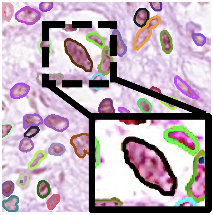
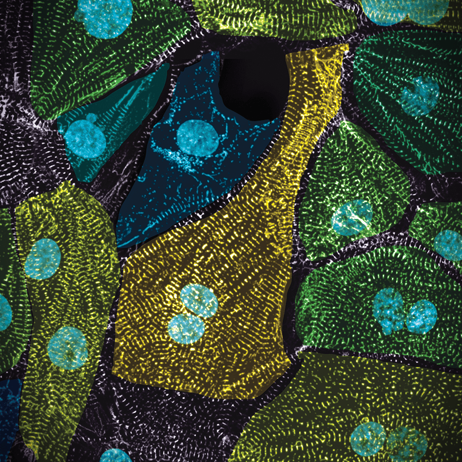
We work side by side with immunologists, neuroscientists, and cardiovascular and cell biologists. When off-the-shelf software can't answer their question, we build the analysis that can: tracking thousands of migrating immune cells, mapping whole-organ vasculature, quantifying tissue change after stroke, building an integrated stem-cell model. Every analysis is validated against what the biology demands, which makes these partnerships our methods' hardest and most honest test.
Selected work
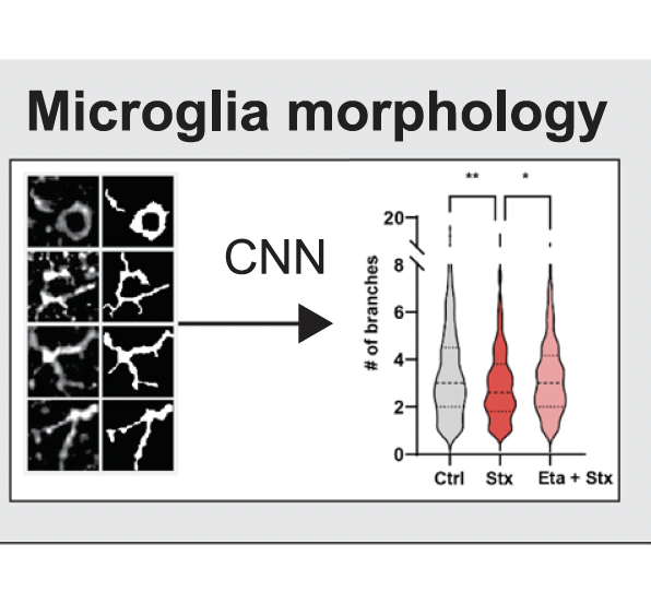
Integrated hiPS cell model · Nature '23 NETs after stroke & MI · Nat Cardiovasc Res '24 Brain microvasculature after stroke · ATVB '24 ComplexEye · Nat Commun '23 Uncertainty-aware cell migration · PLOS Comput Biol '26 CD177 neutrophil subtype · Adv Sci '26 Shiga toxin neuroinflammation · J Neuroinflamm '25 Neutrophil migration · J Leukoc Biol '25 Nuclear growth trajectories · Cell Syst '25
02 Efficient AI for bioimaging
High-end AI with lighter models and smaller data, for better science.
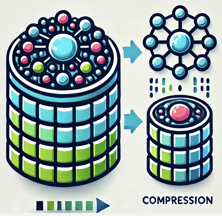
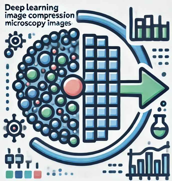
Bioimaging AI only matters if labs can afford to run it and store what it produces. We make networks faster and less energy-hungry without losing accuracy. We also rethink how microscopy images are stored: learned compression, neural representations that encode an image as a compact function, and storage spent only where the biology is. The goal is terabyte-scale imaging that stays analyzable, shareable, and sustainable.
Selected work
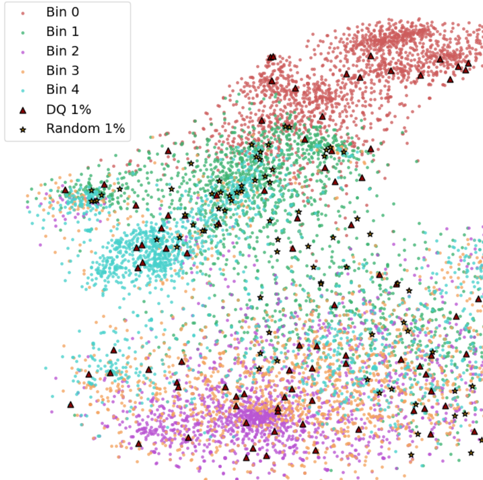
EfficientBioAI · Nat Methods '24 Resource-aware AI · Nat Mach Intell '23 Implicit neural image fields · Nat Comput Sci '25 SGI: 2D Gaussians · CVPR '26 FlowRoI · npj Imaging '26 Learned compression study · Biol Imaging '24
03 AI-ready datasets
Better AI needs better and more data, which requires modern data management.
Most bioimage data never becomes useful for AI. It exists, but it is hard to find, poorly documented, or never quality-checked. We release datasets and benchmarks designed for machine learning from day one, and build the tools to organize, curate, and quality-control images at scale. For us, data release and data management go hand in hand: AI-readiness starts long before publication.
Tools & benchmarks
BioFile Finder · Nat Methods '26 AutoQC-Bench · npj Imaging '25 BIABench · arXiv '26
Released datasets
hiPS cell image atlas · Nature '23 Light-sheet vasculature · Cell Rep Methods '23 Immune cell migration · Nat Commun '23
04 Agents & foundation models
What will bioimage analysis look like in five years?
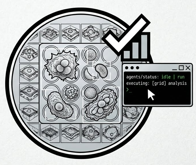
Foundation models and multimodal LLMs are changing what analysis software can be. Instead of chaining tools by hand, a researcher describes the goal, and an agent plans, runs, and checks the analysis. We develop such agents. Their ceiling is set by two things: stronger generalist models, and better specialized tools and data for them to call. So we keep building robust, well-validated tools, from light-sheet image correction to vessel analysis.
Agents & models
MLLMs for bioimage analysis · Nat Methods '24 Agentic-J · arXiv '26 Orochi · NeurIPS '25
Tools for agents (and people)
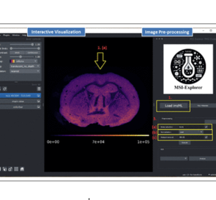
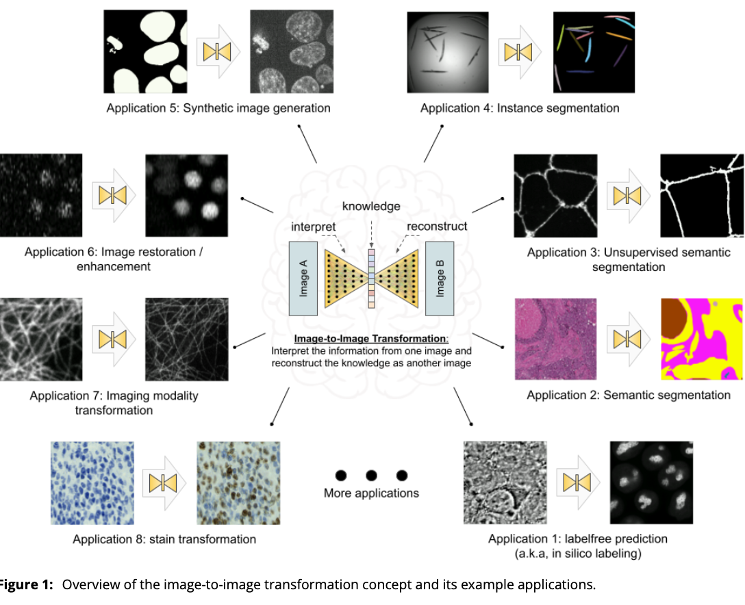
MMV_Im2Im · GigaScience '24 MSI-Explorer · Anal Chem '25 Leonardo light-sheet correction · preprint '25 Vessel analysis · Cell Rep Methods '23
05 Foundations & frontiers
The hard questions underneath every analysis.
Beyond our core themes, we pursue the questions that decide whether bioimaging AI can be trusted and pushed further.
Metric pitfalls · Nat Methods '24 Application-appropriate validation · Nat Methods '23 Risk of manual annotations · ICCVW '23
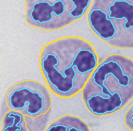
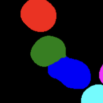
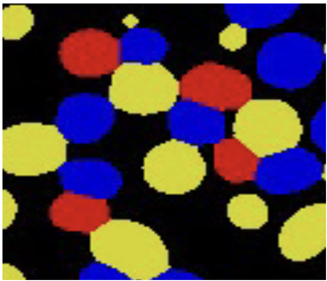
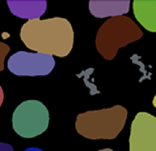
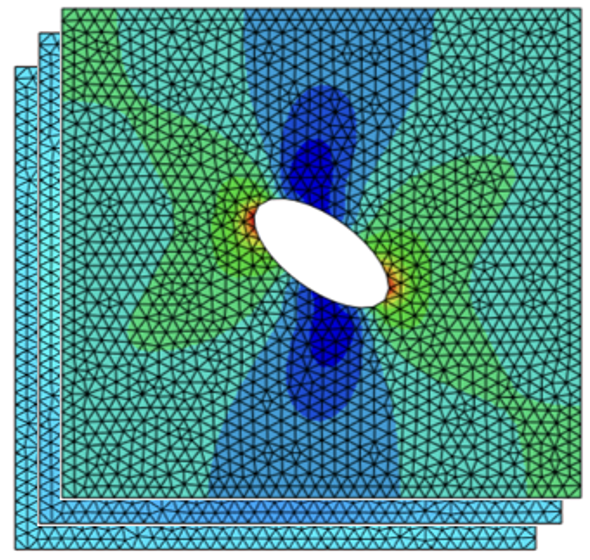
Cell instance boundaries · IEEE TMI Four Color Theorem for segmentation · ICML '25
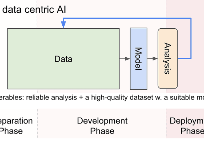
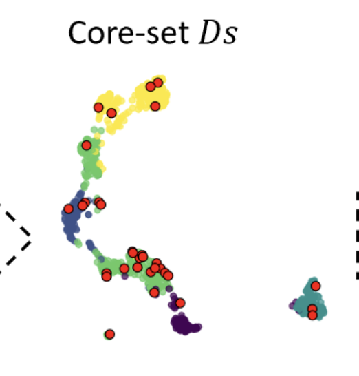
A data-centric lens · npj Imaging '25 Data efficiency & transfer · BIBM '25 OOD detection · BIBM '25
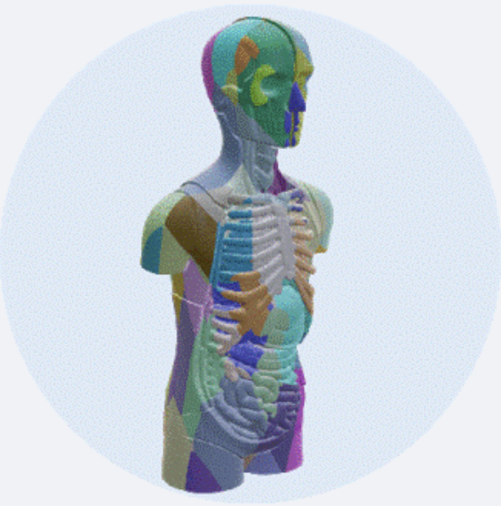
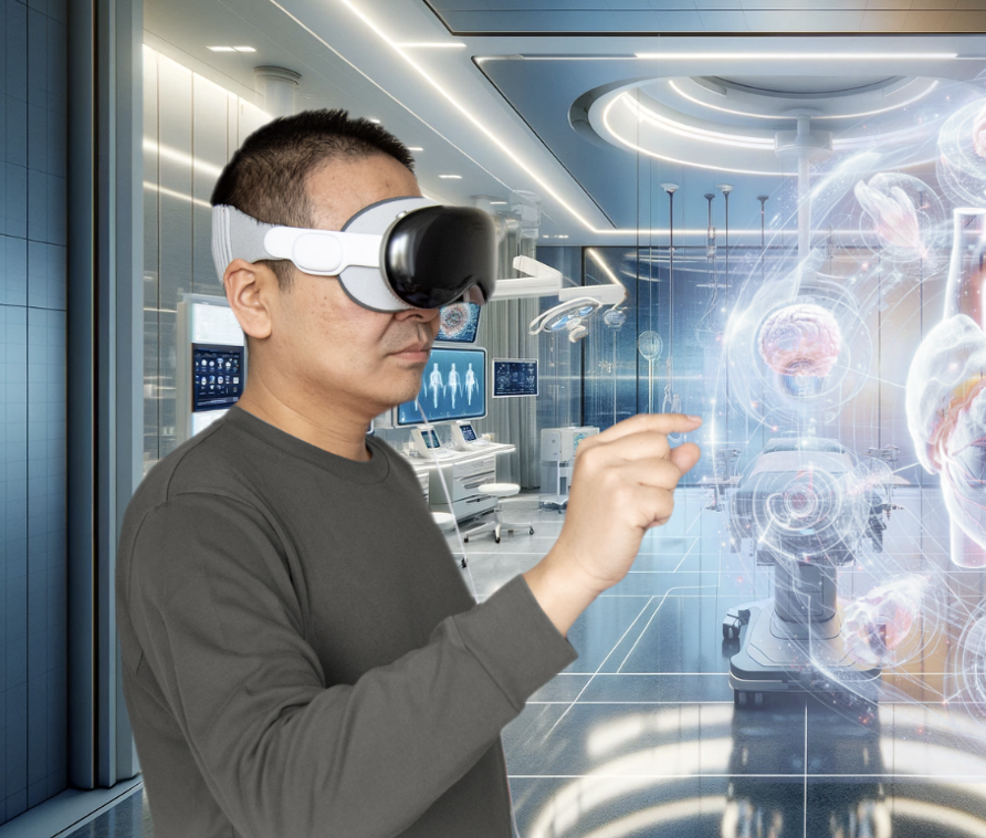
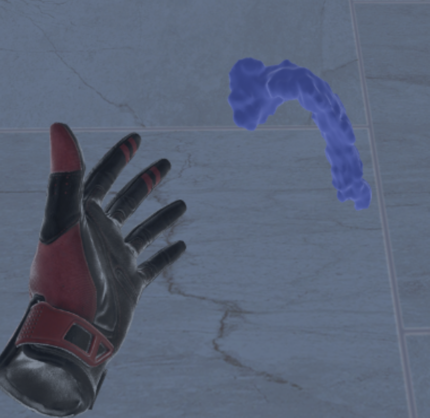
MedShapeNet · Biomed Eng '24 Apple Vision Pro · JMIR Serious Games '24 Multisensory XR · J Imaging Inform Med '24
Outlook Multimodal AI
Microscopy shows where and when; molecules tell us what. We want both in one model.
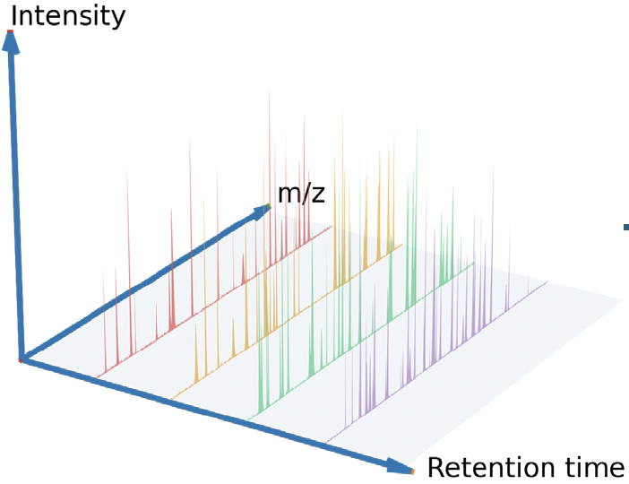
Microscopy captures structure and dynamics; mass spectrometry imaging and other omics capture biochemistry. Learning across these modalities opens biomedical research in four dimensions: quantity, type, location, and time. This is where we're heading, and ISAS, an analytical-sciences institute, is a natural place to do it.
First steps beyond images
MS1 pretraining for proteomics · ACM BCB '26 INFL: federated learning for omics · arXiv '26 Our agents · see 04
2026

2025

2024
2023
2021
2020

2019

2018

2017

2016
2015
2014